Direction Ear Pull for Tympanic Membrane Reading
Vestibular disorders are quite common, and largely considered idiopathic. Information technology was estimated that up to 35% of the U.Southward. population to a higher place 40 years of age has vestibular dysfunction. It affects women upwardly to three times more frequently than men (Neuhauser et al., 2005; 2007). Some of the more known types of vestibular impairment, are BPPV (benign positional paroxysmal vertigo), Meniere's illness, vestibular migraines, idiopathic dizziness, cervicogenic dizziness, vestibular neuritis, auto sickness, boat sickness, etc. Unfortunately, considering these illnesses do not have an officially known etiology, proper testing may not exist. Agrawal et al. (2009) estimated that only 30% of patients with vestibular dysfunction would be diagnosed with the common tests which are used today. Further, the likelihood of acquiring one of the above problems increment with historic period, and is also greatly associated with diabetes mellitus as well as peripheral neuropathic disorders.
From 2001 through 2004, 35.four% of US adults anile forty years and older (69 million Americans) had vestibular dysfunction. – Agrawal et al., 2009
Although vestibular disorders are common and oft disabling, they remain hard to diagnose and care for. – Lewis, 2015
The point of this commodity is not to repeat the well-known, but rather to reveal the lesser known however common relationship between craniocervical dysfunction and the vestibular system. Naturally, this approach should be used when infections, tumors etc. have been excluded, i.e. when the condition is considered "idiopathic". However, permit u.s. first briefly discuss how the vestibular system works.
NB: I have been planning to write this article for a while, but unfortunately I am very busy. Therefore it must be somewhat shorter (abbreviated) than priorly planned. I may fill in some gaps after. Similar simply more detailed information and associations between ear-dysfunction and TMJ / neck associations can be institute in my tinnitus commodity.
The vestibular organisation and its role
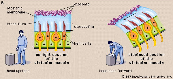
The vestibular portion of the inner ear, likewise known as the labyrinth, consists of two otolith organs: The utricle and sacculus, which annals linear movements (inductive, posterior, lateral, medial, up, downwards), and iii semicircular canals which register rotational movements (yaw, pitch, curl), which are called the superior, posterior and horizontal semicircular canals.
Fig. 1: Vestibular system

The otolith organs consist of a otolith crystals, the otolithic membrane and the stereocilia pilus sensors. The otolith crystals are fastened to the otolithic membrane, as showed below, and they move in accordance with gravitational forces as well as momentum. If someone pushes you frontwards, for example, the otolith & hair sensors would be pulled backwards, creating electrical signals translated from the trunk'due south position and movement to your encephalon and thus allowing precise counter motion being organized in the cerebellum.
The sacculus register vertical (longitudinal) and lateral (coronal) movements, and the utricle register forwards, backwards (sagittal) and lateral movements. Thus they both accept a mutual ground in coronal movements, just are unique in perceiving longitudinal and sagittal movements. This is because the sacculus is positioned in a 90˚ bending compared to the flat-lying utricle.
Fig. two: Saccular and utricular move organs
There are iii semicircular canals, as mentioned already. The posterior culvert registers pitch, which is a coronal axis rotation. The inductive canal registers rolls, which is a sagittal axis rotation. And, finally, the lateral canal registers yaw, which is a longitudinal axis rotational motility.
The pilus sensors in the semicircular canals are stimulated a little differently than the otolith organs. All of the vestibular chambers are filled with endolymphatic fluids, simply like in the cochlea, however the semicircular canals register move of this fluid. During motility, the endolymphatic menstruation volition move the crista hair cells and thus create signals according to which movements are occurring. These signals are then sent to the cerebrum for estimation. The endolymphatic fluid is produced past the stria vascularis, which is the outer wall of the cochlear canal, as well as by dark cells within the labyrinth, located in the sacculus and utricle. It is as well regulated by the endolymphatic sac, as well by the tympanic plexus. Improper endolymphatic regulation volition influence balance, which we will discuss soon.
The stria vascularis and vestibular nighttime cells are the two principal structures responsible for endolymph secretion, and possess many similarities. The characteristics of these structures are the basis for regulation of inner-ear homeostasis – Ciuman, 2009
Fig. 3: Semicircular motion organs

Craniocervical factors that bear on the vestibular system
Several factors, in addition to the patently noted vestibulocochlear nerves (CN eight), may touch on the vestibular system. Several studies bear witness that intracranial hypertension (ICH), sudden intracranial hypotension (SIH), intermittent ischemia, venous congestion and nerve pinch syndromes may dramatically touch on function and estimation of the vestibular chambers. Posture, muscle function and stress levels are all imperative factors to consider when treating vestibular dysfunction.
Thus, this article will expand upon these topics and explain the mechanism of dysfunction also as how i may approach treatment.
Intracranial pressures / Venous congestion
Intracranial pressures are intrinsically related to vestibular function. The reason for this is that endolymphatic fluid levels are regulated through intracranial pressure (ICP) and proper drainage. The cerebral venous system does not have valves, as in the rest of the body, and is thus more sensitive to flow aberrancy. Thus, low pressure level may cause vertigo, as may high pressures (Larsen 2018b, Soler 1998, Friedman 2014, Schievink 1996, 2000). Longstanding ICH may lead to dural perforation (peculiarly in susceptible patients, such equally those with Ehler-danlos syndrome (EDS)) with concomitant CSF leakage and sudden intracranial hypotension (Higgins 2015). Mokri (2002) showed that patient swho had their proven CSF leak fixed, developed hypertension, paradoxically suggesting that the 2 are associated. Farther, longstanding ICH may also cause [secondary] endolymphatic hydrops (Ranieri 2017), a known pathology associated with Meniere's disease, hearing loss and vertigo.
Fig. 4: Proximity of the IJV and CN9-12 to the transverse and styloid processes
Fig. 5: Interscalene triangle & costoclavicular space

The most common cause of idiopathic intracranial hypertension is poor posture. I have named it biomechanical intracranial hypertension (Larsen 2018b). The reason for this, is that droopy shoulders may oft lead to pinch of the distal subclavian artery (SA) (weak and tight scalenes may also). Distal SA obstruction will force blood redirection toward the carotid and vertebral arteries, thus saturating the cerebrovascular arrangement. As a second but perhaps more important problem, is that hinge-neck postures as I have described in many of my articles (atlas, migraine), may crusade blockage of the internal jugular vein, which is the primary venous drainage from the cerebrum. This may occur as the vein is blocked betwixt the styloid process of the cranium and the transverse process of the C1 (Larsen 2018b, Gweon 2011, Dashti 2012, Higgins 2015), and/or wrapped effectually the transverse process (TP) (Flanagan 2014) every bit the patient hyperextends their caput in posture. Jayaraman et al. showed that extracranial IJV obstruction was coincidentally nowadays in 33% of came into their section for angiography (the latter existence negative, and the patients were deemed healthy), indicating that its prevalence is much higher than anticipated. Either way, drainage volition be impaired, and the pressure will build up. More so, if the subclavian artery is obstructed besides. Increased inlet yet obstructed outlet is a recipe for intracranial hypertension, and may outcome in many maladies, some of which being endolymphatic hydrops, vertigo, chronic fatigue, migraine, tinnitus, hearing loss, vision impairment or even blindness (Corbett 1982, Jayaraman 2012).
In many circumstances, the IJV obstacle may be unilateral. Some practitioners may argue that unilateral obstruction is not a big problem, as compensatory drainage occurs through the contralateral sinus and IJV. Still, studies prove that besides unilateral obstruction may still be clearly symptomatic (Cumurciuc 2005; Hyun-Ah Kim 2008; Thandra 2015; Zhou 2017).
Fig. 6: Compression of the internal jugular vein between the SP and C1 TP. Source: Gweon et al. 2011

Fig. 7: Centric MRI: Large forrard atlanto-occipital subluxation of the C1 in a patient with longstanding vestibular dysfunction and ear "fullness"

Fig. eight: Sagittal MRI: Conspicuous frontwards subluxation

Endolymphatic fluid is tuckered out from the ear via the vein of the vestibular aqueduct, and into the sigmoid sinus. As stated, there are no venous valves in this system and thus adequate pressures are needed to maintain fluid balances. If the venous outlet is blocked of the organization is saturated, especially the start-mentioned or both in tandem, endolymphatic drainage volition be greatly dumb and may even retrogade. This leads to longstanding pressure level buildups within the vestibular as well every bit cochlear chambers, with mentioned sequelae of hydrops etc. A common surgical intervention is to remove the endolymphatic sac, a regulative organ for endolymphatic fluid. However information technology is my impression that endolymphatic "overproduction" from the endolymphatic sac is extremely unlikely to crusade such massive amounts of fluid that hydrops occurs. An utterly blocked outlet, potentially with retrogade period is a much more likely culprit.
Fig. ix: Inner ear'southward venous drainage

Information technology has been documented that ICH and SIH may often be an occult disorder, where lumbar puncture opening pressures (LPOP) are normal despite clear symptoms of ICH. Thus one may not solely rely on LPOP for diagnosis of ICH and SIH. MRI criteria for SIH and ICH must exist evaluated thoroughly for each case. MR or CT venography for evaluation of venous integrity should be performed (Higgins 2015, 2017; Larsen 2018b; Mokri 1997; Schievink 1998).
Generally, the problem may be ameliorated past improving cervical and scapular postures, every bit to decompress the IJV and SA. If the atlas has subluxed significantly forrard, strengthening exercises that prevents anterior translation of the C1 will be done. The almost important exercises are those for the longus capitis and levator scapulae.
Migraine-associated vertigo has become a well-recognized disease entity diagnosed based on a clinical history of recurrent vertigo attacks unexplained by other key or peripheral otologic abnormalities, which occurs in the patient with a history of migraine headaches. – Cha, 2010
The inner ear fluids play a major role in the cochlear and vestibular physiology by the transmission of the mechanical stimulus to the hair cells, on the i paw, and by the transduction of this signal to a nerve potential, on the other hand. – Ferrary & Sterkers, 1998
It has too been shown that, the distal portion of the endolymphatic sac, although it may exist large in size may non accept any discernable confluent expanse within it and that it may also overlap the sigmoid sinus (Friberg et al, 1988). There is a thickening of the dura mater effectually the endolymphatic sac which extends to the sigmoid sinus, beyond the end of the sac itself – Locke, 2008
The endolymphatic sac responds to endolymph volume disturbance, showing opposite responses to book increases and decreases. Although prove is all the same limited, the endolymphatic sac appears to act every bit a "bidirectional overflow" system. While book disturbances originating from out-of-balance transport processes anywhere in the labyrinth may exist corrected by the sac, dysfunction of the sac itself is probable to have a substantial effect on endolymph condition – Salt, 2001
Vertebrobasilar insufficiency
Another relatively mutual cause of vestibular dysfunction, yet often one that is occult (hidden), is positional vertebrobasilar insufficiency. It is well known that vertebral artery injuries may upshot in firsthand vertigo. It is less known, however, that intermittent obstruction of the VAs may crusade chronic nonetheless periodic vertigo in absence of concrete VA injury. It may besides cause migraines, vision impairment, etc.
Fig. 10: Vertebral avenue course and segments

The vertebral arteries ascend from the subclavian artery, runs through the transverse foramen betwixt C6-C1, and then enters the cranium to course the basilar artery. The basilar artery supplies the cerebellum, so connects to the posterior circle of Willis (Cow). However, information technology as well supplies the ear via the labyrinthine arteries. It is very common that one of the arteries are underdeveloped (hypoplasia), where the correct avenue is usually smaller than the left i (left dominant VA) (Pellerito & Polak 2012). The labyrinthine arteries carry a small-scale nevertheless crucial blood supply to the inner ear, and it has been documented that the inner ear is very sensitive to ischemia (Nuttall 1999b; Wangemann 2002b). Further, intermittent cerebral ischemia may too lead to vertigo on its own, as dizziness is a known pseudostroke symptom (Tehrani 2014).
Fig. 11: Inner ear'due south arterial supply

It has been shown many times that biomechanical obstruction of the vertebral arteries may occur. It is commonly seen in patients with thoracic outlet syndrome, as the scalenus anticus may compress the VA if information technology branches off the posterior side of the subclavian artery (Powers 1961). It may also get compressed betwixt the scalenus anticus and the longus colli, at the scalenovertebral bending (Kojima 1985). Osteophytic evolution inside the vertebral foramen, or even laterally protruding disc herniations may as well compress the arteries. At that place have been reports of migraine and transient blindness occurring with rotation, where a tight scalenus anticus have ben plant every bit culprit with dynamic angiographic examinations (Sell 1994, Saxton 1998). Positional blockage of the VA has also been found with ultrasound doppler examinations in the DeKleyn'due south position, despite angiography being normal (Arnetoli 1989). Nevertheless, dynamic examinations of the VA is rarely performed, and the patient volition be told that their hemodynamics (blood period) are normal as the pathology is intermittent, positional and occult.
Typical symptoms of TOS are pain between the shoulderblades, arm hurting, carpal tunnel syndrome, radial neuralgia, migraines, cervix hurting, breast pain, animate pain, and more than (Larsen 2018b; Selmonosky 1982, 2002, 2008)
Thoracic outlet syndrome may be ameliorated by strengthening the scalenus anticus and medius carefully, along with postural changes. Bow hunter'south syndrome may somewhat be ameliorated by strengthening the suboccipitals, every bit they prevent excessive C1-2 movement. However, fixation may be necessary in farthermost circumstances. Disc herniations and/or osteophytic evolution inside the transverse foramen may be surgically removed. In such case, a significant reject in systolic speeds will be noted upon doppler ultrasound of the vertebral artery (V2) between one foramen to the next (if a lesion is present).
BPPV can occur secondary to diverse other conditions including viral neurolabyrinthitis, Meniere'south disease, and vertebrobasilar ischemia [137]. In Meniere's disease, information technology has been suggested that hydropic distension or rupture damages the otolithic apparatus, leading to the release of otoconia droppings which migrate to the semicircular canals where they may event in BPPV [138]. Although BPPV is one of the most common causes of vertigo, it is non the only disorder associated with recurrent vertigo. Meniere's disease, migraines, vertebrobasilar insufficiency, and panic disorder are also characterized by recurrent episodes. Caution in diagnosis BPPV is warranted every bit other conditions can present with BPPV-like symptoms. For example, fundamental vestibular disorders can requite ascension to positional nystagmus, which can be mistaken for BPPV. Such lesions tin can arise from posterior fossa such every bit from modest cerebellar strokes [131]. In inner ear pathologies which may cause vestibulopathies, such as perilymph fistulas, nystagmus may exist enhanced past Dix-Hallpike testing. Furthermore, equally spontaneous nystagmus associated with a perilymph fistula improves, nystagmus toward the afflicted ear in the down position may misdirect the examiner from the initial pathological trigger of the symptoms. – Parham, 2014
This theory is supported by the strong correlation of hydrops with Meniere attacks, the finding that autoregulation of cochlear blood flow is impaired in the hydropic ear, and studies demonstrating that symptoms and signs in people and in animal models vary with conditions that modify perfusion pressure in the inner ear. Induction of Meniere attacks in animate being models requires both hydrops and a mechanism that reduces perfusion pressure level, such as epinephrine injection or caput dependency. There is a strong clinical association betwixt Meniere attacks and disorders that increment the risk for cerebrovascular ischemia, such as migraine. – Foster & Breeze, 2013
Thoracic Outlet Syndrome (TOS) causes dizziness because of positional compression of the vertebral artery with resultant symptoms of vertebrobasilary insufficiency. Compression of 7,C8,and T1 fretfulness fibers is responsible for the neck pain. – Selmonosky, 2007
The cases of 17 patients with vertigo, tinnitus, deafness, supraclavicular bruit, and a macerated radial pulse are reported. All the patients had an anomalous vertebral artery. All had subclavian-vertebral arteriograms preoperatively. Each patient showed an anomaly of the vertebral artery organisation which allowed intermittent pinch of either the origin or cervical form of the artery. The compression was commonly aggravated past rotation or hyperextension of the neck. In about cases, the vertebral artery arose at the level of the thyrocervical trunk and the compression was relieved by section of the scalenus anticus muscle and by partitioning of the inferior thyroid avenue. – Powers et al., 1961
We report a patient who developed occasional vertigo when turning his head to the correct side. Apoplexy of the right vertebral artery occurred at the narrowed "scalenovertebral angle" with this rotational head motion. This triangular tunnel consisted of the hypertrophied ligament of the longus colli muscle and the anterior scalene musculus. – Kojima et al., 1985
Rotation-induced vertebrobasilar artery hypoperfusion causes transient ischemic attacks (TIAs), affecting the cerebellum, brainstem and spinal cord. When these symptoms occur transiently due to head movement, compression of the vertebral artery by an extraluminal lesion should be suspected. Cervical spondylotic spurs and anterior scalene musculus or deep cervical fascia are amidst the factors which can compress the vertebral avenue. – Dadsetan & Skerhut, 1989
Rotational positioning of the caput showed vertebral obstruction in one direction, and unobstructed filling of the vessel when the head was turned to the contrary side. Fifteen patients showed rotational vertebral artery occlusion. The site of obstacle occurred at the origin of the vertebralartery or cephalad to the level of C5. 2 patients had bilateral fascial band obstacle, one patient had left only, and the remaining 10 were obstructed on the correct side. An inductive scalenotomy was done with preservation of the phrenic nerve. We were more impressed with the deep cervical fascia as the cause of intermittent rotational obstruction rather than the anterior scalene musculus. Just 2 patients showed unequivocal poststenotic dilatation as evidence of severe anterior scalene muscle compression. The obstructing extra-luminal fascia was quite dumbo, fibrotic and ofttimes completely encircling the artery. – Hardin & Poser, 1963
Subclavian steal symptoms presents secondary to arterial insufficiency, created by a retrograde menstruation that "steals" blood from the brain circulation, more specifically from the basilar artery via the vertebral artery. Classically information technology presents with neurological symptoms from the posterior encephalon and cerebellum [iv,six]. Decreased catamenia over the basilar artery gives rise to symptoms like lightheadedness, ataxia, vertigo, dizziness, confusion, headache, nystagmus, hearing loss, presyncope and syncope, visual disturbances, focal seizures, and in extremely rare cases, expiry [6–ten]. However the vast majority of patients are asymptomatic and rarely require any intervention [3,5,xi]. – Alcocer et al., 2013
This article describes migraine without aura since babyhood in a patient with bilateral cervical ribs. In addition to usual migraine triggers, symptoms were triggered past neck extension and by arm abduction and external rotation; paresthesias and pain preceded migraine triggered by arm and neck movement. Suspected thoracic outlet syndrome was confirmed by high-resolution bilateral magnetic resonance imaging (MRI) and magnetic resonance angiography (MRA) of the brachial plexus. An unsuspected aberrant right subclavian artery was compressed within the scalene triangle. Left scalenectomy and rib resection confirmed the MRI and MRA findings; the scalene triangle contents were decompressed, and migraine symptoms subsequently resolved. – Saxton et al., 1999
Thoracicoutlet syndrome (TOS) refers to the pinch of the neurovascular bundle within thethoracicoutlet. Cases are classified by primary etiology-arterial,neurogenic, or venous. In addition to the typical symptoms of arm swelling and paresthesias, headaches have been reported equally a potential symptom of TOS. In this report, nosotros describe a patient with debilitating migraines, which were consistently preceded by unilateral arm swelling. Resolution of symptoms occurred only subsequentlythoracicoutlet decompression. Patients with migraines and concomitant swelling and/or paresthesias, especially related to provocative arm maneuvers, should be considered a possible atypical presentation of TOS and evaluated in more detail. – Chahwala et al., 2017
It is as well noteworthy that the hypertrophied and contracted anterior scalenus muscle exerts a stiff although intermittent pinch of the vertebral artery, causing in severe TOS diverse symptoms that are very characteristic of vertebrobasilary insufficiency. – Silva & Selmonosky, 2011
Reports of transient blindness resulting from this condition are even more rare. The authors draw the case of a middle-aged woman who presented with transient blindness when she turned her head excessively to the left. She as well exhibited other less severe brainstem symptoms. Arteriography demonstrated occlusion of the left vertebral artery merely when her head was rotated to the left. Surgical exploration revealed entrapment of the left vertebral artery past a tight anterior scalene muscle, release of which resulted in complete resolution of her symptoms. – Sell et al., 1994
Nerve entrapment syndromes
It is commonly thought that vestibular dysfunction is attributed to vestibular nerve impairment. "Vestibular nerve neuritis" is a common yet generic and oft fake diagnosis. It may too be chosen "idiopathic dizziness". It must exist understood and considered the tympanic plexus is also greatly involved in regulating vestibulocochlear function. Many nerves contribute to the tympanic plexus, which may likewise exist irritated in the neck and temporomandibular joint. This is why patients with neck and jaw disorders have a high prevalence of vestibular co-dysfunction, in my experience.
The tympanic plexus is formed by several nerves, namely the glossopharyngeal nerve, the facial nerve, and sympathetic fibers coming from the internal carotid plexus. Notwithstanding, it also communicates with the trigeminal nerve via the otic and pterygopalatine ganglia too as the vagus nerve via the glossophargyneal nervus. It supplies the mucosa of the middle ear, the mastoid cells, the auditory tube, and parotid glands. – Barral & Croibier,Transmission Therapy for the Cranial Nerves, 2009
Fig. 12: Tympanic plexus contribution

The tympanic plexus is involved in regulation of endolymphatic fluid levels, opening of the eustachian tube and thus besides tympanic chamber pressures, the mucous membranes as well as mastoid air cells. When the fretfulness which contribute to the tympanic plexus are compromised, e.one thousand. entrapped, this may cause faulty signalling to occur within the ear's nervous network. The nearly mutual nerves to become compromised are the vagus nerve, sympathetic plexus and trigeminal nerves.
The glossopharyngeal nerve may get entrapped between the styloglossus and stylohyoid muscles. Further, it anastomosis with the vagus nerve. The vagus nervus transmits betwixt the scalenus anticus and clavicular caput of the sternocleidomastoid muscles, and may go entrapped hither. It may also go compressed betwixt the cranial styloid process and the C1 transverse procedure, every bit it passes between these structures in upwardly to 66% of the population (Kim & Ledwitz-Rigby, 2014). One is especially predisposed to this problem with poor craniocervical postures and/or forrad subluxation of the atlas (encounter my atlas article for more information). Vagus nerve entrapment will also oftentimes cause eustachian tube dysfunction, explaining why many patients with vestibular disorders also experience "fullness" of the ears or clogged" ears (Park et al. 2012). This resolves when the entrapment has been dealt with. Other symptoms include burning tongue syndrome, pharynx pain, "lumpy" throat, chronic or periodic idiopathic dry cough, excess hiccups (phrenic nerve), tinnitus, dysphagia (swallowing difficulty), itchy throat, ear hurting, etc.
If the compression of the neurovascular parcel occurs at the atlantostyloid interval, this indicates that the atlas has come forward. It can likewise be a positional problem (Larsen 2018c). The patient must cease hinging at the neck and tucking their chin and follow the guidelines which I outlined to a higher place also as. in my atlas article. If in that location is myofascial entrapment of the glossopharyngeal nerve within styloglossus and stylohyoid, the patient must stop clenching their hyoid muscles in posture. If it is vagus nerve entrapment between the SCM and scalenus anticus, strengthening of these muscles will be appropriate intervention. Over again, proper posture must also be maintained throughout the twenty-four hour period.
Fig. 13: Oromandibular grade of the glossopharyngeal nerve

Fig. 14: Cervical course of the vagus nerve

The sympathetic plexus contribute straight to the tympanic plexus. It may become entrapped between the cervical alar fascia and the longus colli & longus capitis muscles, causing lengthened symptoms of dysautonomia such equally vestibular and vision harm, tinnitus, itchy ears, ear clicking, eustachian tube dysfunction, etc. The superior cervical and stellate ganglia of the sympathetic chain has been implicated in Meniere's disease (Franz 1998, 2007, Kang et al. 2005, Raj 2007), and I have personally (anecdotally) confirmed this association. Strengthening of the longus colli and longus capitis along with postural correctives are the main intervention for this problem.
Fig. 15: The sympathetic plexus transmits between the alar fascia and longii muscles

The trigeminal nerve (auriculotemporal branch) may become compressed betwixt the mandibular condyle and TMJ fossa, hence its implication in TMD. It has been well documented that patients with TMD may struggle with symptoms of trigeminal neuralgia as well as vestibular impairment (Levandowski 2008). Further, several researchers accept shown that the trigeminal nerve directly innervates the vestibular and cochlear chambers (Liu & Xu 2016; Vass 1998, 2001, 2004; Shore 2000) and that its stimulation may arm-twist vestibular dysfunction such every bit nystagmus (Marano 2005). The trigeminal nerve besides innervates the tensor tympani muscle, whose dysfunction has been implicated in Meniere's disease. Also, the facial nerve innervates the stapedius musculus. Tenotomy of the stapedius and tensor tympani muscles has been shown to profoundly reduce vertigo and tinnitus (Franz 2003). However, are the muscles really to arraign, or are they symptoms of compromised nerves? My impression is that the latter is true. In addition to vertigo and dizziness, trigeminal nervus compression may cause tinnitus, tremendous temple headaches, eye pain, palatine pain, molar aches, cheek sensitivity, ear pain, etc.
Auriculotemporal nerve pinch is a mechanical phenomenon and must be treated as such. Frontwards move of the mandible in posture (see video below) also equally strengthening of the pterygoid muscles will ameliorate the situation as it causes mandibular protraction and thus TMJ decompression. The patient must also be assessed for whether or non they clench their suprahyoid muscles for neck stability (video below).
Fig. 16: Auriculotemporal nervus course posterior to the mandibular condyle

In support of the neural theory, the Barré-Liéou syndrome or posterior sympathetic syndrome has often been mentioned. Its mechanism is, yet, uncertain and has been discredited, allowing misuse of this eponym [16]. This socalled posterior sympathetic syndrome has recently been challenged, as vestibulocochlear symptoms can exist explained by the "anterior sympathetic" arrangement comprising the superior cervical ganglion and its postganglionic neurons, with branches to the eustachian tube and inner ear [17]. Overall, prove suggests that disturbances of the cervical spine tin can exist associated with Ménière's disease symptoms and that their adequate direction tin lessen inner-ear symptoms. – Franz & Anderson, 2007
The consequence of novocain block on vertigo of Meniere'south disease. I have had the satisfaction of abruptly terminating two cases of Meniere's disease during acute severe attacks past means of a procaine block. 1 case occurred at the Wembley Hospital. The patient was wheeled into the theatre lying curled up airsickness, with nystagmus, pallor and sweating. five ml. of procaine were injected in the stellate ganglion and inside three minutes the patient had recovered sufficiently as to walk dorsum unaided to the ward. –Garnett Passe, Sympathectomy in Relation to Meniere'south Disease, Nervus Deafness and Tinnitus
Superior cervical ganglion neurons projection to the dilator pupillae musculus of the iris to control pupil dilation. Ocular blood flow is controlled both via direct autonomic influences on the vasculature of the optic nerve, choroid, ciliary body, and iris, as well equally via indirect influences on retinal blood flow. – McDougal & Gamlin, 2015
It is more often than not believed that the cause of Meniere'southward disease is related to autonomic dysfunction (Hilger', 1950; Beickert', 1953; Watanabe10, 1955; Hisaki', 1960; Williams», 1965). The positive charge per unit showing a response of either the sympathetic hyperreactor or sympathetic hyporeactor blazon in the cases with Meniere'south disease and aural vertigo was 79% and 87% respectively in the acute stage when nystagmus was present. – Uemura et al., 1972
MANDIBULAR joint neuralgia (Costen's syndrome), first reported by Costen in 1934,1 is accepted by otorhinolaryngologists and members of the dental profession every bit a definite clinical entity. Information technology should be considered in every differential diagnosis of recurring facial hurting. – Beyes & Teich, 1952
Co-ordinate to the author'south hypothesis, the lack of posterior support of the alveolar ridge led to mandibular vertical acme loss which caused a slipping backward of the condyles over the articular disc thus resulting in TMJ discal damage, erosion of the glenoid fossa bone, pinch of the Eustachian tubes and tympanic plates and consequent impingement of the auriculotemporal nervus (ATN), which runs on the postero-medial attribute of the TMJ capsule, and chorda tympani nerve4 . – Paparo et al., 2008
Normal spacing between the roof of the glenoid fossa of the temporal bone and the condyle of the mandible should exist approximately three mm to back up the disk between them. The retrodiskal tissues originate from the distal portion of the glenoid fossa and are inserted into the posterior portion of the disk. This tissue contains a matrix of claret vessels and fretfulness, particularly fibers of the auriculotemporal nervus, cranial nervus V, an afferent branch of the trigeminal nerve. If this space is bereft or reduced or restricted and the condylar head grows posterosuperiorly or is iatrogenically repositioned posteriorly or posterosuperiorly, the condyle volition pinch this tissue and usually the result will be pain. – Sims & Stack, 2007
Our clinical work suggests that the auriculotemporal (AT) nervus, a co-operative of the mandibular nerve, the largest of the three divisions of the trigeminal nerve, plays a critical part in TMD sequelae. The AT nerve provides the somatosensory fibers that supply the articulation, the centre ear, and the temporal region. By projecting fibers toward the otic ganglion, the AT nerve establishes an important bridge to the sympathetic system. As information technology courses posteriorly to the condylar head of the TMJ, compression, injury or irritation of the AT nerve tin atomic number 82 to significant neurologic and neuro-muscular disorders, including Tourette's syndrome,Torticolli, gait or balance disorders and Parkinson's disease. Subsequent irritation and compression of the AT nerve tin can occur, with associated parasthesia, pain and discomfort. Symptoms can be local and specific (eastward.yard., TMD), as well every bit varied and systemic (due east.chiliad., neurologic, dystonic and neuro-muscular disorders, including tremors, muscle spasms leading to dumb and bad-mannered positional control of the head, hands, other extremities, speech impairment, incontinence, impaired sleep, associated depressive symptomatology). – Demerjian et al., 2011
Anatomical relationships between the auriculotemporal nerve and the muscles of mastication, temporomandibular articulation, and surrounding vessels in the area of the infratemporal fossa create favourable conditions for entrapment syndromes. Entrapment of the auriculotemporal nerve plays a role in the pathogenesis of temporomandibular joint pain syndromes, headaches, as well as pain symptoms or paraesthesias within the external acoustic meatus and auricle. Komarnitki et al., 2012
The syndrome of symptoms (Tabular array 1) equally first described by Costen, an American otolaryngologist, was discussed. Costen attributed the symptoms to temporomandibular joint dysfunction consistent upon mandibular overclosure with distal condylar deportation. He assumed that the displaced condyle might lead to whatever of the following: Compression of the eustachian tube, erosion of the glenoid fossa or tympanic plate, force per unit area on the chorda tympani, or force per unit area on the primary trunk of the auriculotemporal nerve. – Clarke, 1962
Sectioning of the tensor tympani and stapedius muscle tendons significantly reduced the frequency and intensity of vertigo and improved both the functional profile and tinnitus. – Franz et al. 2003
Suboccipital dysfunction
The suboccipital muscles are a unique muscle group with a very high density of receptor cells. They are the almost local stabilizers of the craniocervical junction, and take a muscle fiber configuration that allows them to motility the caput in all various directions. The reason for this, is that the suboccipitals synchronize the head with the eyes' movements. If you palpate them while moving your eyes effectually, you'll experience them pop upward nether your fingers, according to your ocular move. Very interesting, indeed!
When they are adequately dysfunctional, meaning extremely weak, the patient may first experience dizziness during motion such every bit in the machine or boat sickness. Equally the dysfunction progresses, the dizziness may go more pronounced. Weakness of the suboccipitals may as well cause tremendous headaches and may mimic migraines. Information technology may also cause excessive movement of the atlanto-occipital and atlantoaxial joints when overly lax / weak. I never recommend massaging these muscles; only strengthening. It is imperative to empathise that weak muscles feel tight and are painful, and that this will exacerbate with continuous stretching, yet permanently resolve with proper strengthening. You tin can detect exercises for these muscles in my atlas article and/or on my youtube channel.
Fig. 16: Suboccipital muscles

The distribution and organisation of spindles inside the muscle and their arrangement was studied. The spindle density of superior oblique muscle was establish to be 190, that of inferior oblique was 242 and the rectus capitis posterior independent 98 spindles per gram of muscle. – Kulkarni et al., 2001
Muscle spindle density is extremely loftier in the deep muscles of the human neck. – Liu et al., 2003
Stress levels
Everyone knows that stress is an amplifying factor. Withal, a common tendency of people who are very stressed, scared, anxious etc., is that they hold their breaths and brace their bodies. I phone call this phenomenon "global involuntary clenching strategy" (GICS), where the patient is more or less constantly creating valsalva maneuver due to inability to handle stress. Constant valsalva volition cause intracranial hypertension as well as musculus imbalances, and may therefore be directly linked to vestibular dysfunctions and not merely an amplifying factor. I will not elaborate upon this topic here, but rather you can read about it in my GICS article.
Within the poorly understood mechanisms implicated in the aetiology of beneficial paroxysmal positional vertigo (BPPV), the results of this trial provide clinical evidence of a potential function of emotional stress connected to adverse life events every bit a trigger of otoconial dysfunction. High levels of anxiety, depression and somatization were recorded and considered psychogenic precursors of BPPV, thus emphasizing the role of psychological distress in precipitating peripheral vestibular disorders. Therefore, appraisal of life stress and psychological attitudes may have potential implications in the clinical assessment of this labyrinthine vertigo and its frequent relapses. – Monzani et al. 2006
The postural common denominator
The importance of posture is a very controversial topic these days. Still, I recollect it is counterintuitive to reject posture. Clearly, the fashion nosotros hold ourselves greatly bear on how we use our muscles. It's not coincidental that patients with poor postures often have very painful muscles. However, we need to know the specifics of what expert posture really is. I think the main reason that posture remains controversial, is the lack of very important nuances. Cues to "straighten the back" or "lower the shoulders" are detrimental every bit they discourage natural postural habits (Larsen 2018a, 2018b).
Loss of lumbar lordosis is causing patients to clench their abs in posture, inhibiting them from animate properly. Stress exacerbates the poor breathing pattern, and sets them on course in an evil circle. Moreover, low of the clavicles, which is very mutual, will promote swivel-neck postures as well as compression of the costoclavicular space (thoracic outlet syndrome) (Swift 1984, Telford 1948, Larsen 2018b). As I touched upon earlier, costoclavicular space compression (CCS) may shrink the subclavian artery, redirecting more than blood into the head. Hinge-cervix postures may block the internal jugular vein, thus potentially creating a huge imbalance between arterial inlet and venous outlet, resulting in craniovascular saturation and intracranial hypertension.
Although many "sub-diagnoses" tin can be made, I sincerely believe that poor posture and stress are the main and fundamental causes of vestibular disorders. That said, once the malady has progressed and matured, both posture and proper corrective rehabilitation is required to opposite the trouble.
Case case
Beneath is the instance of a 45 year old adult female who had sudden onset of vertigo. She has a history of several whiplash accidents and has suffered from both jaw and neck pain for many years, yet has been active and working throughout this time. She was initially diagnosed with vestibular neuritis, just as time went by she started to develop other cognitive dysfunctions such as dysarthria (speech difficulty), confusion, disorientation, facial numbness and left-sided transient facial hemiparesis (facial paralysis, too known as Bell'southward palsy or seventh nerve palsy). She was nor able to say "R" nor hold a normal conversation, or even sign papers. She was admitted to the hospital and several examinations were performed, yet no concrete pathology was found. Her MRIs were deemed "normal", and she was sent home.
When she visited my role, she presented with a significantly poor posture, with very distended external jugular veins, indicative of intracranial hypertension. I set her in proper posture, and inside two minutes she was able to pronounce "R" and look me in the eyes. Her colleague, which assisted her, confirmed that her ability to interact had improved on those minutes. This indicates that at that place is biomechanical pinch of the IJVs (Larsen 2018c), or else she would not improve by postural alteration lonely.
Afterwards, going through the MRIs (I received these at her second visit), to my surprise, I saw articulate signs of intracranial hypotension (low pressure), not hypertension. Either way, non so "normal" findings after all, as priorly stated by the hospital. I was likewise able to detect stenosis of the left IJV, which was caused by forrard subluxation of the atlas. Ultrasound besides showed hypoperfusion in the left IJV and hyperperfusion in the right IJV, besides equally rounded systolic peaks of the left ICA and vertebral artery, which is uniform with her stroke-like symptoms equally well as the MRI images. As stated before, long-term hypertension can cause SIH (sudden intracranial hypotension) (Higgins 2015). This is. what happened here. Her lumbar puncture opening pressure was. 11,5 cm H2O (ref: > 10). Clearly, with a blocked vein, i would expect higher levels than xi,5, unless in that location is indeed a leak. Such a sudden leak in the following of chronic venous congestion would explicate the sudden onset of vestibular and cognitive impairment.
half dozen weeks after the postural correction and initiation of neck and jaw rehabilitation, she is once again able to agree a normal conversation, pronounce "R", solve math puzzles, sign papers, her memory has returned, and concluding but not least, has a 95% reduction in dizziness. The CSF leak heals on its own once the IJV blockage resolved. This has also been documented by Higgins (2015). I presume that's besides what happened here.
Fig. 17: Signs of SIH

Fig. 18: Forward subluxation of the left atlanto-occipital joint, explaining fig. 19

Fig. nineteen: Left IJV stenosed at the atlantostyloid interval

Fig. 20: Hypoperfusion (poor drainage) in the left IJV

Fig. 21: Relatively normal right IJV. There is a slight only insignificant stenosis hither every bit well.

Fig. 22: High-force per unit area "splash-catamenia" in right IJV in try to recoup for left stenosis

Case #2 – Positionally conditioned
Beneath is a 38 year one-time woman with proven intracranial hypertension without known crusade ("idiopathic"). She suffered whiplash in the early 2000. Her lumbar puncture opening pressure was 50cm Water in 2003, and she was diagnosed with idiopathic intracranial hypertension. A shunt to drain out the excess CSF was installed, normalizing the ICP to around 15cm H2O. Her headache was somewhat ameliorated still many symptoms persisted, and she was ultimately not able to keep working. She notwithstanding had tremendous cervix pain, perceived intracranial pressure level with eye pain, and chronic headaches. Around 2015 it was postulated that it may exist related to venous congestion, then she underwent CT venography, however this examination was negative. Then, in 2018 she sought a seated MRI examination abroad, which did non explain her symptoms either, except for indications of atlantoaxial instability.
Fig. 23: Falsely normal atlantostyloid interval and unobstructed IJVs considering the patient didn't lie in a mode which simulated her natural posture.

In person, the patient one time again presents with very poor neck and shoulder posture, suggesting biomechanical IJV stenosis equally a possible etiology of her problems. Due to the atlantoaxial instability, I examined her vertberal arteries with rotation (with ultrasound), just this examination was unremarkable. Thus excluding arterial insufficiency as the cause of her remaining symptoms. Examination of the MRIs however evidence that there was still flattening of the posterior eye (global flattening), and concave partially empty sella (pituitary compression), which indicate the presence of ICH. Farther, in opposition with the lying CTV, the upright MRI showed obliteration of the left internal jugular vein, and partial obstruction of the right 1. Doppler confirmed these findings, and too showed slowed systolic upstrokes. At present, why would at that place even so be bear witness of ICH if the CSF is drained via the shunt? Because excess CSF is merely a secondary symptom! The main problem here has been occult venous congestion since the problem'southward genesis. Thus the CSF backlog is a mere sequela of internal jugular vein stenosis. I fix this patient in proper posture, which has reduced her symptoms considerably. We are also rehabilitating her cervix and shoulder problems. Every bit well-nigh patients with a history of whiplash and associated disorders (WAD), this patient also had TOS (thoracic outlet syndrome).
Fig. 24: Indicator of ICH (Osborn's brain, 2017)

Fig. 25: Indicator of ICH (Osborn's encephalon, 2017)

Fig. 26: Obstruction of IJVs in upright position with poor posture

Fig. 27: Atlantoaxial hypermobility

Fig. 28: Hyperperfusion (compensatory period) right IJV

Fig. 29: Very slight perfusion in the left IJV with "proficient posture"

Fig. xxx: All the same, zero left IJV perfusion when lying in her natural posture ("bad posture" or "BP")

Fig. 31: Quite aberrant left ICA waveforms, notwithstanding the right ICA was more or less normal, uniform with the left IJV obliteration

This case is a good example of how patients with poor posture may take falsely negative CTV or MRV results when searching fore extracranial stenosis, if they are not lying down in their natural posture. As mentioned, extracranial stenosis is a little known yet common problem (Jayaraman 2012).
Treatment & detection
It takes time to get to the point of chronic vestibular dysfunction. The patient must be aware that the treatment is rough and that it is not a quick fix. First and foremost he or she must resolve their poor posture. The patient needs to larn to properly curvation their lower backs in posture and to finish bracing, i.east. to terminate holding their breath, clenching their abs and creating valsalva habitually. Moreover, they demand to maintain a "long neck" position and enhance their shoulders in posture, until the superior bending of the scapulae are leveled with the T2 vertebra (Osar 2012, Larsen 2018b, Larsen 2018c).
Poor posture for sixteen hours and and so 2 minutes of cosmetic exercise is a futile strategy and needs not exist attempted. It is fundamentally necessary to practice proper posture AND supplement this with proper cosmetic rehabilitation of impaired soft tissues. The most of import and commonly impaired muscles in vestibular disorders, are the longus capitis and colli, scalenus medius and anticus, suboccipitals, levator scapulae and trapezius muscles. Rehabilitation should be started with very, very light stimulus two-3 times per week. Ambitious or even moderate strengthening may cause tremendous flareup of their symptoms. This is harmless, simply unnecessary. Start piece of cake, and gradually build the muscles up.
Intracranial pressures: Lumbar puncture opening pressures are known to be unreliable. The practitioner should non pass up SIH or ICH based on a negative LPOP. Schievink (1998, 2000) which is one of the foremost researchers on SIH, states that LPOP is not needed to diagnose SIH, but rather, the physician should be guided by the appearance of the cerebral MRIs equally well as the patient'southward clinical symptoms. Imaging signs for ICH are such equally, merely non limited to: Flattened posterior globes with or without optic nerve bulging, optic nerve sheath distension, undulated optic nerve, [partially] empty sella, pons flattening, obliteration of the basal cisterns and cerebral sinuses, chiari, or "tight brain" advent. Signs for SIH are such equally, just not express to: CSF-depleted tractus opticus, distended transverse and sagittal sinuses, < 50˚ pontomesencephalic angle, enlarged pituitary, brain sagging, chiari, basal cistern effacing, elongated midbrain, lateral ventricle narrowing, mamillopontine distance < 5,5 mm (Osborn's brain, 2017).
Detection of venous blockage can be washed through MRV or CTV (5=venography), but it is important that the patient lies with their natural cervical and scapular position during the examination. A seated examination is not necessary. Any arterial insufficiency, more often than not implying rotational insufficiency of the vertebral arteries, can exist detected with a doppler browse with and without rotation of the neck. The patient will be scanned in the DeKleyn's provocative position. However, information technology is important that the physician scans the V3 segment (the supraatlantal segment) and not the V2, in instance of Bow hunter's syndrome. In either case, a significant systolic decline with signal loss, tardus parvus waveform, or potential absence of flow volition be noted. Confirmation should be done with dynamic MRA or CTA (A=angiography). Detection of stenosis via MRV or CTV (venography) is underutilized and under-appreciated, according to Higgins 2004. Almost probable because IJV stenosis at the craniocervical junction is non a well-known problem. Similarly, there are few practitioners measuring the vertebral arteries with rotation of the neck.
As for rehabilitation of atlanto-occipital instability and/or nerve entrapment syndromes, these topics are too large to echo one time more, when I've written in lengths about them in other articles, such as my neck pain, thoracic outlet syndrome, temporomandibular joint disorder and atlas joint instability articles. Confer with these for rehabilitative protocols. The main telescopic of this article is to show WHY the neck and TMJ may greatly touch on the vestibular arrangement.
In conclusion
If no concrete findings or causes for vestibular impairment are found, such equally tumors or similar, then craniocervical and TMJ interest should be considered as a potential crusade. Poor postures may crusade chronic intracranial venous congestion with subsequent intracranial hypertension. Information technology may also promote thoracic outlet syndrome, which as been implicated in intermittent vertebrobasilar insufficiency. Both of these issues are known etiologies of chronic vestibular dysfunction. Further, the nerves from the neck and jaw contribute to the tympanic plexus, and may thus be implicated in dysregulation of the middle and inner ear structures, equally well as eustachian tube. Cervix and jaw disorders are treatable and fifty-fifty curable with the right approaches.
References:
- Neuhauser HK, von Brevern M, et al. Epidemiology of vestibular vertigo: A neurotologic survey of the general population. Neurology. 2005;65(6):898–904.
- Neuhauser HK. Epidemiology of vertigo. Curr Opin Neurol. 2007;20(one):40–46.
- Agrawal Y, Carey JP, Della CC, et al. Disorders of Residue and Vestibular Role in The states Adults. Arch Intern Med. 2009;169(10):938-944. doi:10.1001/archinternmed.2009.66.
- Lewis RF. Advances in the diagnosis and treatment of vestibular disorders: psychophysics and prosthetics. J Neurosci. 2015;35(13):5089-5096. doi:10.1523/JNEUROSCI.3922-14.2015
- Ciuman RR. Stria vascularis and vestibular dark cells: characterisation of main structures responsible for inner-ear homeostasis, and their pathophysiological relations. J Laryngol Otol. 2009 Feb;123(2):151-62. doi: 10.1017/S0022215108002624. Epub 2008 Jun 23. PMID: 18570690.
- Soler D, Cox T, Bullock P, et al. Diagnosis and management of benign intracranial hypertension. Athenaeum of Disease in Childhood 1998;78:89-94.
- Friedman D. (2014) The pseudotumor cerebri syndrome. Neurol Clin 32: 363–396.
- Schievink WI, Meyer FB, Atkinson JLD. et al. Spontaneous spinal cerebrospinal fluid leaks and intracranial hypotension. J Neurosurg. 1996;84:598-6058613851
- Schievink WI, Ebersold MJ, Atkinson JLD. Roller-coaster headache due to spinal cerebrospinal fluid leak. Lancet. 1996;347:14098637363Google ScholarCrossref
- Schievink WI. Spontaneous spinal cerebrospinal fluid leaks. Neurosurg Focus. 2000;9:one-nine
- Mokri B. Intracranial Hypertension After Handling of Spontaneous Cerebrospinal Fluid Leaks. Mayo Clin Proc. 2002;77:1241-1246
- Higgins N, Trivedi R, Greenwood R, Pickard J. Brain slump caused by jugular venous stenoses treated by stenting: a hypothesis to link spontaneous intracranial hypotension with idiopathic intracranial hypertension. J Neurol Surg Rep. 2015 Jul;76(1):e188–e193. doi: x.1055/s-0035-1555015
- Ranieri A, Cavaliere Thou, Sicignano S, Falco P, Cautiero F, De Simone R. Endolymphatic hydrops in idiopathic intracranial hypertension: prevalence and clinical outcome later on lumbar puncture. Preliminary data. Neurol Sci. 2017 May;38(Suppl one):193-196. doi: 10.1007/s10072-017-2895-8. PMID: 28527079.
- Gweon HM, Chung TS, Suh SH. Evaluation of the Cause of Internal Jugular Vein Obstruction on Caput and Neck Contrast Enhanced 3D MR Angiography Using Contrast Enhanced Computed Tomography. JKSMRM 15:41-47(2011)
- Dashti SR, Nakaji P, Hu YC, Frei DF, Abla AA, Yao T, et al. Styloidogenic jugular venous pinch syndrome: diagnosis and handling: case written report. Neurosurgery. 2012 Mar;lxx(3):E795-9. doi: 10.1227/NEU.0b013e3182333859
- Larsen K. Occult intracranial hypertension equally a sequela of biomechanical internal jugular vein stenosis: A case report. Anaesth Hurting & Intensive Care 2018;22(two)
- Flanagan MF. The Office of the Craniocervical Junction in Craniospinal Hydrodynamics and Neurodegenerative Conditions. Neurol Res Int. 2015;2015:794829. doi:x.1155/2015/794829
- Corbett JJ, Savino PJ, Thompson HS, Kansu T, Schatz NJ, Orr LS, Hopson D. Visual loss in pseudotumor cerebri. Follow-up of 57 patients from 5 to 41 years and a contour of xiv patients with permanent severe visual loss. Arch Neurol. 1982 Aug;39(8):461-74. doi: 10.1001/archneur.1982.00510200003001. PMID: 7103794.
- Jayaraman MV, Boxerman JL, Davis LM, Haas RA, Rogg JM. Incidence of extrinsic compression of the internal jugular vein in unselected patients undergoing CT angiography. AJNR Am J Neuroradiol. 2012 Aug;33(vii):1247-50. doi: x.3174/ajnr.A2953. Epub 2012 Feb 9. PMID: 22322614.
- Cumurciuc R, Crassard I, Sarov M, Valade D, Bousser MG. Headache as the merely neurological sign of cerebral venous thrombosis: a series of 17 cases. J Neurol Neurosurg Psychiatry. 2005 Aug;76(8):1084-vii. doi: ten.1136/jnnp.2004.056275. PMID: 16024884; PMCID: PMC1739763.
- Kim HA, Sohn SI, Lee H. Cerebral venous thrombosis mimicking acute unilateral vestibulopathy. Neurol Sci. 2008 Feb;29(1):41-3. doi: 10.1007/s10072-008-0858-9. Epub 2008 Apr 1. PMID: 18379740.
- Karaman E, Saritzali 1000, Cansiz H. A instance of increased intracranial pressure after unilateral modified radical neck autopsy. Am J Otolaryngol. 2009 Jul-Aug;30(four):261-3. doi: 10.1016/j.amjoto.2008.04.007. Epub 2008 Sep 21. PMID: 19563938.
- Thandra A, Jun B, Chuquilin M. Papilloedema and Increased Intracranial Pressure equally a Effect of Unilateral Jugular Vein Thrombosis. Neuroophthalmology. 2015 Jul 15;39(4):179-182. doi: 10.3109/01658107.2015.1044541. PMID: 27928352; PMCID: PMC5123161.
- Zhou D, Meng R, Zhang X, Guo 50, Li S, Wu Westward, Duan J, Song H, Ding Y, Ji Ten. Intracranial hypertension induced by internal jugular vein stenosis can be resolved by stenting. Eur J Neurol. 2018 February;25(2):365-e13. doi: 10.1111/ene.13512. Epub 2017 Dec 7. PMID: 29114973.
- Higgins JNP, Pickard JD, Lever AML. Chronic fatigue syndrome and idiopathic intracranial hypertension: Different manifestations of the aforementioned disorder of intracranial pressure? Med Hypotheses. 2017 Aug;105:vi-9. doi: 10.1016/j.mehy.2017.06.014. Epub 2017 Jun 24. PMID: 28735654.
- Mokri B, Piepgras DG, Miller GM: Syndrome of orthostatic headaches and diffuse pachymeningeal gadolinium enhancement. Mayo Clin Proc 1997, 72:400–413.
- Schievink WI, Morreale VM, Atkinson JL, et al.: Surgical handling of spontaneous spinal cerebrospinal fluid leaks. J Neurosurg 1998, 88:243–246.
- Cha YH. Migraine-associated vertigo: diagnosis and treatment. Semin Neurol. 2010 Apr;30(ii):167-74. doi: 10.1055/s-0030-1249225. Epub 2010 Mar 29. PMID: 20352586; PMCID: PMC5682200.
- Ferrary E, Sterkers O. Mechanisms of endolymph secretion. Kidney Int Suppl. 1998 Apr;65:S98-103. PMID: 9551441.
- Locke RR. Beefcake of the Transmastoid Endolymphatic Sac Decompression in the Management of Ménière'south Disease. The University of Glasgow Faculty of Biomedical and Life Sciences Apr 2008
- Table salt AN. Regulation of Endolymphatic Fluid Volume. THE VESTIBULAR LABYRINTH IN Wellness AND Disease October 2001. Volume942, Issue1. p306-312
- Pellerito J, Polak J. Introduction to Vascular Ultrasonography sixth Edition, 2012; Elsevier publishing.
- Nuttall AL. Sound-Induced Cochlear Ischemia/Hypoxia as a Mechanism of Hearing Loss. Dissonance & health. 1999b;two:17–32. [PubMed] [Google Scholar]
- Wangemann P. Cochlear blood flow regulation. Adv Otorhinolaryngol. 2002b;59:51–57.
- Saber Tehrani Equally, Kattah JC, Mantokoudis G, Pula JH, Nair D, Rush A, et al. Small strokes causing severe vertigo: Frequency of false-negative mris and nonlacunar mechanisms. Neurology. 2014;83:169–173.
- Powers SR Jr, Drislane TM, Nevins Southward. Intermittent vertebral avenue compression; a new syndrome. Surgery. 1961 Feb;49:257-64.
- Kojima N, Tamaki N, Fujita K, et al: Vertebral artery apoplexy at the narrowed "scalenovertebral bending": mechanical vertebral occlusion in the distal starting time portion. Neurosurgery 16:672–674, 1985
- Sell JJ, Rael JR, Orrison WW. Rotational vertebrobasilar insufficiency as a component of thoracic outlet syndrome resulting in transient blindness. Case written report. J Neurosurg 1994; 81: 617–9
- Saxton EH, Miller TQ, Collins JD. Migraine complicated past brachial plexopathy as displayed by MRI and MRA: abnormal subclavian artery and cervical ribs. J Natl Med Assoc. 1999 Jun; 91(half dozen): 333–341.
- Selmonosky, C.A. Byrd, R. Blood, C. Blanc, J.S. Useful triad for diagnosing the crusade of chest hurting. S. Med. J. 1981, 74, 947-949.
- Selmonosky C.A, Silva R.P. The diagnosis of thoracic outlet syndrome. Myths and facts. Revista Chilena de Cirugia 2008, sixty, 255-261.
- Selmonosky, C.A. Thoracic outlet syndrome. The missing link in the diagnosis of non-coronary chest hurting. Italian Journal of Cardiology 2008, 9, 217S.
- Parham M. Benign Paroxysmal Positional Vertigo: An Integrated Perspective. Advances in Otolaryngology. Book 2014 |Commodity ID 792635 | https://doi.org/10.1155/2014/792635
- Foster CA, Breeze RE. The Meniere assail: an ischemia/reperfusion disorder of inner ear sensory tissues. Med Hypotheses. 2013 Dec;81(half dozen):1108-15. doi: 10.1016/j.mehy.2013.10.015. Epub 2013 Oct 22. PMID: 24199949.
- Dadsetan, M.R., Skerhut, H.E. Rotational vertebrobasilar insufficiency secondary to vertebral avenue occlusion from gristly band of the longus coli muscle. Neuroradiology 1990, 32, 514-515.
- Hardin CA, Poser CM. Rotational Obstruction of the Vertebral Avenue Due to Redundancy and Extraluminal Cervical Fascial Bands. Annals of Surgery: July 1963 – Book 158 – Consequence 1 – ppg 133-137
- Alcocer F, David Chiliad, Goodman R, Jain SK, David Southward. A forgotten vascular disease with important clinical implications. Subclavian steal syndrome. Am J Case Rep. 2013;14:58-62. doi:10.12659/AJCR.883808
- Chahwala 5, Tashiro J, Li X, Baqai A, Rey J, Robinson HR. Ann Vasc Surg. 2017 February;39:285.e5-285.e8. doi: 10.1016/j.avsg.2016.05.109. Epub 2016 Aug 13. Venous Thoracic Outlet Syndrome as a Cause of Intractable Migraines.
- Kim Pow, Ledwitz-Rigby F. Variations in the anatomical structures surrounding the transverse process of the atlas (C1) in 15 cadavers. Experimental Biology 2014 Coming together Abstracts V28, consequence S1. April 2014. (915.14).
- Park MS, Lee HY, Kang HM, Ryu EW, Lee SK, Yeo SG. Clinical manifestations of aural fullness. Yonsei Med J. 2012;53(five):985-991. doi:10.3349/ymj.2012.53.5.985
- Franz B, Anderson C. The Potential Office of Joint Injury and Eustachian Tube Dysfunction in the Genesis of Secondary Ménière'due south Disease. International Tinnitus Journal, Vol. 13, No. 2, 132–137 (2007)
- Franz B, Collis-Brownish Thou, Altidis P, et al. Cervical trauma and tinnitus. Int Tinnitus J 4:31–33, 1998.
- Raj PP. Stellate Ganglion Block. Raj's Practical Management of Pain (Fourth Edition), 2007
Source: https://mskneurology.com/vestibular-impairment-and-its-association-to-the-neck-and-tmj/
0 Response to "Direction Ear Pull for Tympanic Membrane Reading"
Post a Comment